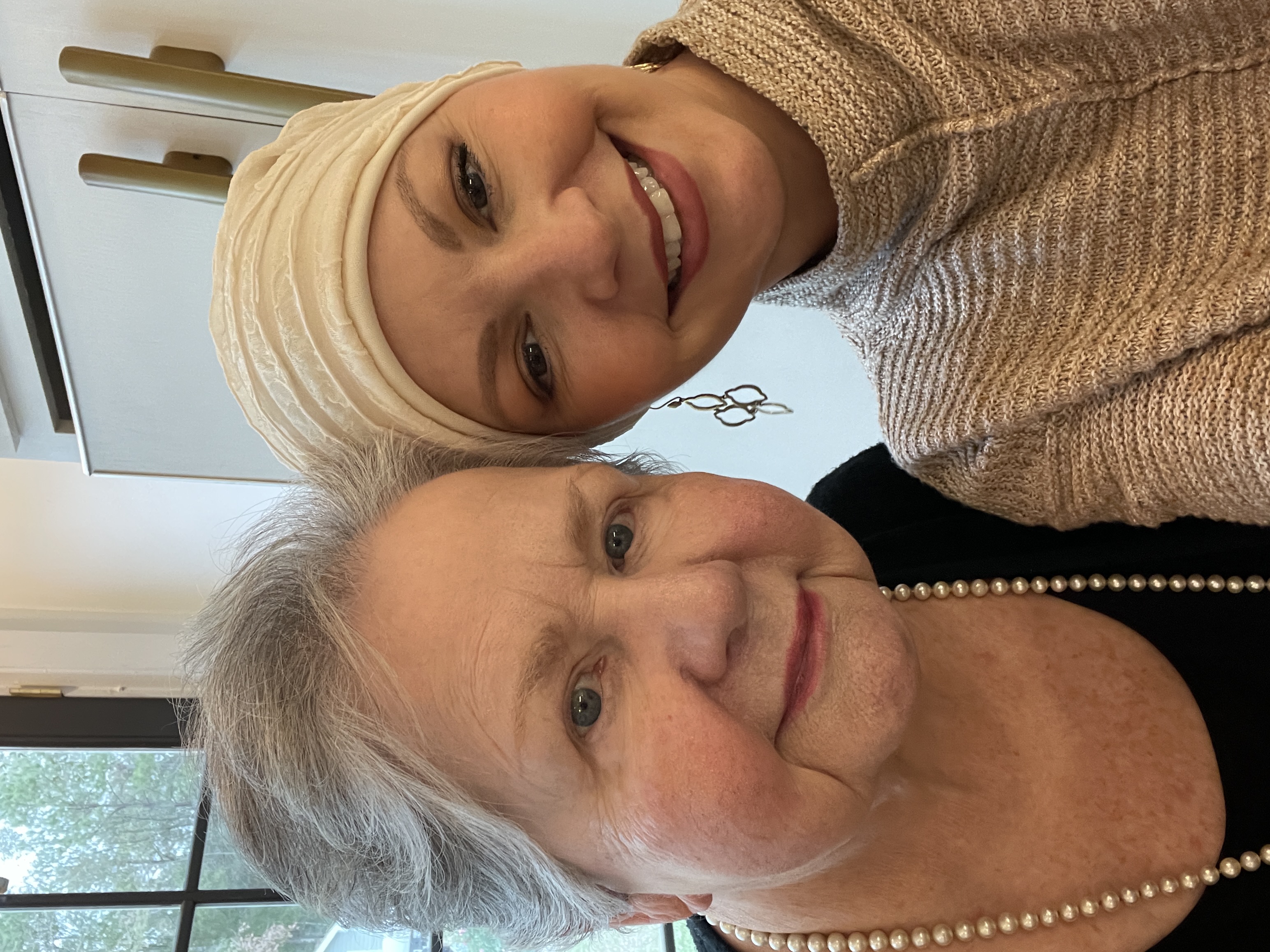
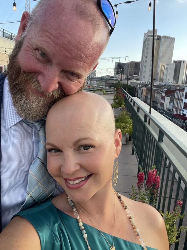
Following a normal mammogram interpretation on February 28th of 2023, I thought I could check off the screening exam for another year and move on. To my surprise, I was diagnosed with malignant lobular carcinoma of the left breast 3 months later. In fact, less than 3 weeks after the normal mammogram, I felt a large mobile mass in my breast. Because of the negative imaging, I assumed the mass was fibrocystic change or perhaps a large cyst that had formed. I allowed several weeks to pass before informally asking the family practice physician I worked with to examine the area which had now begun to pucker the skin at the nipple. My colleague suggested an ultrasound, but I opted to put off the ultrasound and make some lifestyle changes (increase water and decrease caffeine intake) first. Unfortunately, 1 week later, the site began to radiate pain through my left axilla and flank. The diagnostic ultrasound was performed 1 week following the onset of pain, confirming the need for biopsy of multiple masses of the left breast and 1 lymph node of the left axilla. The biopsies were performed 1 week after the diagnostic ultrasound, and the biopsy results confirming breast cancer were discussed with the me 1 week later.
The whirlwind diagnosis and invasion of unsettling feelings created a surreal reality for me at this time. I had relaxed in the comfort of having a normal mammogram screening and being cleared for another year; yet, within weeks, I was plunged into the fear and uncertainty of facing malignant breast cancer. As a nurse, I immediately fell into procedural mode: planning, scheduling appointments, and accessing resources to swiftly move forward with treatment. As a wife and mother, I rehearsed how to share the diagnosis and plan with my family, while also reassuring my loved ones and allaying their fears. As a patient, I began to question the lack of findings on my mammogram and my faith in screening.

Port Placement and Complications
Four days after meeting my treatment team, I had a BardPort® (Bard Access Systems, Inc.) placed by a general surgeon, and chemotherapy was initiated the same day as the port placement. I started chemotherapy 10 days following my diagnosis and 4 days following my initial meeting with the cancer treatment team. Unfortunately, a rare port complication was discovered when the infusion nurse attempted to access the port for my third chemotherapy infusion.
The BardPort® is one of several brands of implantable venous access devices. These devices, introduced into practice in the 1980s, are known by many names and come in a variety of forms. Such names include implantable subcutaneous venous access device, vascular access device, and subcutaneous venous access device.2 All are commonly and simply referred to as a “port” by health care professionals and recipients. Brand names for ports include Mediport®, BardPort®, PowerPort®, and Port-A-Cath®.1 These types of ports are implanted for use in patients requiring long-term intravenous infusions. Such infusion therapy can include biologics, chemotherapy, fluids, hemophilia factor therapy, and intravenous gamma globulin (IVIG).2 The infusions are performed by accessing the port through the thin layer of skin with a specialized needle via the port septum.2
Ports are placed into a subcutaneous pocket created by a surgeon or interventional radiologist under monitored anesthesia care or general anesthesia. The pocket is usually located to the right upper chest, below the clavicle. Incisions are made at the base of the neck and subclavicular to the upper chest. The port is implanted into the subcutaneous pocket and connected to the catheter at the catheter connector site of the port. The small neck incision is accessed to insert the guidewire to the vein, which is punctured for catheter insertion.1 The vein most frequently used is the right subclavian vein (SCV), although the internal jugular vein (IJV) can also be used, as it was in my case. Sutures are then placed at the port’s catheter insertion site, and the incisions are sutured closed. Although there are holes located in the base of the reservoir for suture fixation, this is not standard practice among most practitioners.3 The surgeon who placed my port routinely uses dissolvable sutures with implanted port insertion. According to one study, using non-dissolvable sutures with implanted ports to secure the reservoir to the pectoralis base of the fibrous pocket makes removal of the port “more difficult.”3 However, the authors report that the practice does not lead to an increase in morbidity, cost, or procedure time and would be considered insignificant.3
There are some complications of ports, which include inversion, extravasation, fracture of the catheter, pinching-off of the catheter, infection, and embolism; infection and embolism being the most serious in terms of morbidity and financial burden.4 The reported incidence of port inversion is low (0.2%), according to McNulty et al.3 Another study of 1930 patients by Etezadi and Trerotola5 comparing inversion rates between port types reported an overall rate of 0.9%. Although the incidence is low, the complication of inversion can be significant, resulting in delayed treatment, pain, emotional distress, and need for surgical revision.
Twiddler’s Syndrome
Port inversion is, according to some sources, a direct result of “Twiddler’s syndrome (TS)”.6 The phenomenon of TS was identified and named in 1968 by Bayliss et al,7 with reference to implanted pacemakers. Since its debut, TS has been identified in other implantable devices, such as ports. Twiddler’s syndrome is manipulation of the port beneath the skin resulting in enlargement of the fibrous pocket. In the case of implanted pacemakers, TS can lead to electrode dislocation and result in failure to pace the cardiac rhythm and, in some cases, unintended stimulation of adjacent nerves.7

Port Inversion
My port was placed on May 12, 2023, by a general surgeon. Twenty-two days later, I weeded a garden bed for approximately 1 hour using my right arm crossed over my body. Following this activity, I noticed that I could no longer feel the 3 protrusions marking the surface of the port septum. Instead, the port site felt slightly larger and smooth. On day 42, a Friday, following port implantation, the infusion nurse attempted to access my port unsuccessfully. The markers surrounding the port septum were no longer palpable, and the needle hit the impenetrable metal backing of the port. The infusion nurse determined at that time that the port had inverted. The infusion nurses began preparation to administer my chemotherapy peripherally to avoid delay in treatment. I assumed the inversion had occurred when weeding the garden. I knew the basic anatomy of the BardPort® and was familiar with how it moved within the fibrous pocket. Assuming the port had flipped counterclockwise, I attempted to flip the port over clockwise. By curling my upper torso forward and effectively loosening the skin and soft tissue of the breast/upper chest area, I succeeded in manually rotating my port until I was able to palpate the markers of the septum once again. The infusion nurse was notified, and my port was tested for blood return and flushed successfully with normal saline. Subsequently, I was able to have my chemotherapy administered via the BardPort®, as scheduled.
On day 58, my port inverted once again, after I was in a left side-lying position reading for approximately 1 hour. I flipped the port back to its correct position once again but could not be certain I had rotated the device in the correct direction to avoid creating a kink in the catheter. If the port had now twisted in the same direction a total of 4 times, twice on its own and twice manually, it was possible that the port catheter may have had a kink in it, which could mean the port might not be patent. Not confident that I was palpating a ridge in the tubing rather than a kink, I contacted my oncologist with the concern. I was seen in the surgeon’s outpatient clinic for evaluation prior to my fourth round of chemotherapy, and the port demonstrated acceptable blood return and was successfully flushed. The surgeon reported using dissolvable sutures when the port was implanted. The rare incidence of port inversion may have resulted in the need for surgical revision and possibly tacking with non-dissolvable sutures, had I not been able to successfully flip the port back into position each time. As of day 126, my port has not inverted a third time and is still operational.
In hindsight, I can recall initially being fascinated with the movement of the port under my skin and periodically moving the device laterally, back and forth. Although it is unknown whether my manipulation of the port directly resulted in the inversion, had I been screened for or educated about the possibility of inversion, I would have been less likely to manipulate the port. Additionally, I remained active in the initial weeks following port implantation, so it is possible that my upper body movement led to inversion. Unfortunately, there is a lack of literature devoted to port inversion, likely due in part to the documented low incidence.
Insufficient Research and Patient Education
There is little research on incidence of port inversion with a port that is sutured or tacked in place versus not sutured.3 Studies on the subject do not compare tacking versus not tacking the port in place. Existing predictor tools for TS were not found in searching the literature. Similarly, there is no standard preoperative education regarding TS for patients receiving a port. I was given a patient ID Card containing information on the brand and model of the device for the purpose of informing health care professionals needing to access it.
CONCLUSION
The incidence of port inversion is low and, in my case, the port inversion did not result in a treatment delay or necessitate surgical revision. However, the additional office visit to the surgeon and use of health care resources in messaging between the infusion nurse and myself and the oncology team is a negative outcome of the port inversion. These issues, as well as the personal time and emotional stress I incurred could have been avoided with minor education on activity restriction and avoiding manipulation of the device beneath the skin. Additionally, a predictor or screening tool for patients at higher risk for port inversion would be beneficial in preventing this complication. Studies linking patient activity levels, risks for TS, and education level with incidence of inversion would also be helpful in creating such a tool.
REFERENCES
1. “About your implanted port.” Memorial Sloan Kettering Cancer Center, www.mskcc.org/cancer-care/patient-education/your-implanted-port. Accessed 15 Sept. 2023.
2. “Intravenous (IV) lines, catheters, and ports used in cancer treatment.” American Cancer Society. https://www.cancer.org/cancer/managing-cancer/making-treatment-decisions/tubes-lines-ports-catheters.html. Accessed 15 Sept. 2023.
3. McNulty NJ, Perrich KD, Silas AM, Linville RM, Forauer AR. Implantable subcutaneous venous access devices: is port fixation necessary? A review of 534 cases. Cardiovasc Intervent Radiol. 2010;33(4):751-755. doi.org/10.1007/s00270-009-9758-5
4. Akhter N, Lee L. Utilization and complications of central venous access devices in oncology patients. Curr Oncol. 2021;28(1):367-377. doi:10.3390/curroncol28010039
5. Etezadi V, Trerotola SO. Comparison of inversion (“flipping”) rates among different port designs: a single-center experience. Cardiovasc Intervent Radiol. 2017;40(4):553-559. doi:10.1007/s00270-016-1546-4
6. Ghanchi H, Taka TM, Bernstein JE, Kashyap S, Ananda AK. The unsuccessful twiddler: a case of twiddler’s syndrome without deep brain stimulator lead breakage. Cureus. 2020;12(4):e7786. doi:10.7759/cureus.7786
7. Bayliss CE, Beanlands DS, Baird RJ. The pacemaker-twiddler’s syndrome: a new complication of implantable transvenous pacemakers. Can Med Assoc J. 1965;99(8):371-373.


